

The Uphoff lab is located in the Department of Biochemistry, within the Microbiology and Systems Biology theme. Our central aim is to understand how bacteria adapt to environmental stresses, such as exposure to antibiotics, toxic molecules, and attack by immune cells. Stress adaptation is a multifaceted process that spans scientific fields from molecular biology to population genetics. Biochemical and structural approaches have provided a detailed understanding of the molecular mechanisms involved in cellular stress responses, but it remains an open challenge to unravel the complex interconnections between the myriad intracellular processes, between the cell and its environment, and between the cells in a population. We have developed a toolbox of single-molecule and single-cell fluorescence microscopy methods to address this challenge.
Single-molecule tracking allows us to monitor the movement of individual proteins to understand their functions inside living cells. Using microfluidic chips, we can image thousands of individual bacteria simultaneously and monitor their stress responses and cell fates under precisely controlled treatments over time. To link phenotypic and genetic changes during stress adaptation, we developed a microscopy-based method to detect mutation events in individual cells. We complement microscopy with genetics techniques, quantitative data analysis, mathematical modelling, and computer simulations. Combining these methods allows us to trace bacterial adaptation across enormous spatial and temporal scales, from cell populations down to individual molecular events. Our discoveries have shed light on how bacteria withstand antibiotics, alkylating agents, oxidative stress, and UV light, and have revealed mechanisms of DNA damage repair, gene transcription, and chromosome organisation.
We also collaborate with groups at Oxford and internationally on a range of projects related to genome maintenance in bacteria and eukaryotes.
Single-molecule imaging in live cells
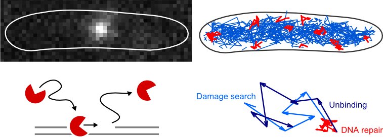
We are applying single-molecule fluorescence microscopy to directly observe individual proteins in living bacteria. One measurement of this type provides a wealth of quantitative information about molecular mechanisms at a single-cell level, such as the abundance and localizations of reaction sites, and the diffusion coefficients and dissociation constants of proteins and their complexes. This approach is very general and powerful, and continues to generate novel insights into the organization of DNA repair pathways, DNA replication, transcription, and chromosome organization.
For example, we used single-molecule tracking to show how the mismatch repair endonuclease MutH finds its target sites in cells. MutH switches from fast diffusion to a slow-search mode near DNA replication forks, allowing it to independently locate hemimethylated GATC sites ahead of any mismatch signal and await activation by MutS-MutL to make the strand incision that triggers repair.
Find more information in these publications: Moores et al. bioRxiv 2026; Stracy et al. Nature Communications 2016, Uphoff et al. DNA Repair 2014; Uphoff et al. PNAS 2013.
Microfluidics for single-cell analysis
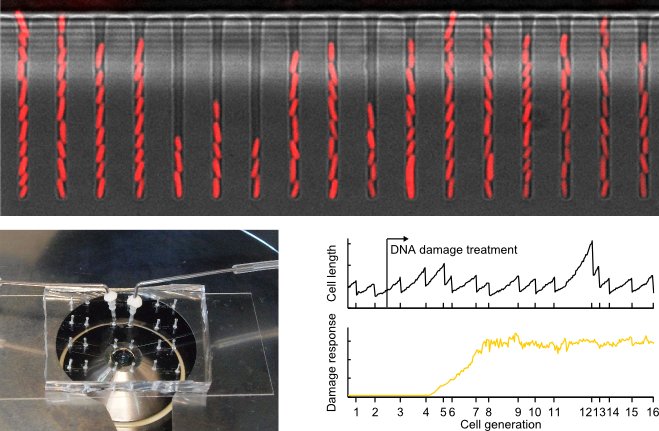 We use microfluidics to investigate how individual cells respond to stress and adapt. The image shows E. coli cells growing inside microfluidic channels. Single cells at the bottom of each channel can be observed continuously over hundreds of generations. This allows us to follow response dynamics and measure phenotypic variation. These experiments inform about the regulation of DNA damage responses and cell fates.
Find out more information in these publications: Uphoff et al. Science 2016, Uphoff PNAS 2018
We use microfluidics to investigate how individual cells respond to stress and adapt. The image shows E. coli cells growing inside microfluidic channels. Single cells at the bottom of each channel can be observed continuously over hundreds of generations. This allows us to follow response dynamics and measure phenotypic variation. These experiments inform about the regulation of DNA damage responses and cell fates.
Find out more information in these publications: Uphoff et al. Science 2016, Uphoff PNAS 2018
How do bacteria regulate stress responses and adapt?
Studying bacterial stress responses at the level of single molecules and single cells has challenged the conventional wisdom of how these processes are regulated and what their functions are. How does a single transcription factor coordinate the response of dozens of genes to protect a bacterial population from oxidative stress? We found that the master regulator OxyR orchestrates its target genes with distinct spatial and temporal dynamics, allowing cells to separate rapid detoxification from longer-term adaptation. Extending this work, we discovered that oxidative stress can drive individual bacteria into chaotic oscillations in gene expression. This is one of the first demonstrations of deterministic chaos in a biological signalling system, and a possible route to generating the phenotypic diversity that helps bacterial populations survive unpredictable environments. We also found that interactions between neighbouring cells shape how the oxidative stress response varies across a population, and that a delay in activating stress responses causes a transient burst of mutations as cells adapt. We are now applying these concepts to understand how heterogeneous stress responses affect antibiotic tolerance.
Find out more in these publications: Choudhary et al. Cell Systems 2024; Choudhary et al. Current Biology 2023; Choudhary et al. Cell Reports 2023; Lagage et al. EMBO Reports 2022.
Tracing the route from phenotypic tolerance to genetic resistance
A central goal in genetics is to understand how mutation rates are regulated by the many genes that act in the creation or prevention of mutations. This is a long-standing challenge, largely because traditional methods for measuring mutagenesis rely on accumulating mutations over time across large cell populations.
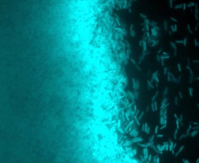
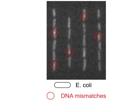
To overcome this limitation, we developed a method to detect mutation events in real time in individual living cells, visualising nascent DNA mismatches (the precursors of mutations) as fluorescent spots within growing cells. This method revealed that mutagenesis in response to DNA alkylatio damage occurs in a distinct pulse shaped by the expression of alternative genome maintenance pathways: mutagenic damage tolerance is required for initial cell survival, before the balance shifts in favour of accurate repair. Cell-to-cell heterogeneity in gene expression was found to modulate these mutation rate dynamics. The approach is generalisable, and showed how mutation rates change in response to antibiotic treatments.
At the molecular level, all biological processes are subject to chance, so even genetically identical cells show random variation in their behaviour. We asked whether such variation affects the formation of mutations in the genome. Revisiting the adaptive response to DNA methylation damage that was discovered in Escherichia coli back in 1977, but using modern super-resolution fluorescence microscopy and single-cell microfluidic imaging, we found that the sensor protein Ada was present at extremely low, and highly variable, copy numbers in unstressed cells. Many cells had none at all, purely by chance. Cells with no copies of Ada failed to sense DNA damage and could not activate the adaptive response, leading to the formation of mutations. This showed that the long-standing dogma that genetic heterogeneity causes phenotypic heterogeneity also holds in reverse: stochastic variation in a cell's capacity to repair its DNA can itself cause genetic change.
Together, these findings suggest that evolutionary adaptation can be driven not by the bulk population, but by subpopulations of cells with elevated mutation rates. We are now extending this work to explore how mutation rate dynamics and heterogeneity, whether stochastic or deterministic in origin, influence the evolution of antibiotic resistance.
Find out more in these publications: Uphoff PNAS 2018; Uphoff et al. Science 2016; Vincent & Uphoff Nucleic Acids Research 2021; Lagage et al. EMBO Reports 2022.
Super-resolution microscopy method development
Super-resolution microscopy by single-molecule localization, such as STORM or PALM, employ photoswitching to image a small subset of molecules at a time while the majority of fluorophores reside in a non-fluorescent state. Thus isolated emitters can then be localized with high precision. The entire set of localizations acquired over the course of a movie forms a super-resolution image. Extension of this approach allows tracking the movement of individual molecules to directly monitor their activities in living cells.
We develop fluorescence microscopes that maximize detection sensitivity, mechanical stability, and flexibility for our different applications. These hardware developments are accompanied by a suite of custom data analysis software that we continuously improve and modify for each project. We also explore ways for fluorescent labelling of proteins inside live cells.
Choosing the right fluorescent label is critical for reliable single-molecule tracking. We directly compared genetically encoded photoactivatable fluorescent proteins with long-lived HaloTag-dye conjugates in live bacteria, and developed methods to robustly quantify tracking data for fluorophores with different photophysical properties, helping researchers choose the best label for their experiment.
Find more information in these publications: Banaz et al. J Phys D 2018; Moores & Uphoff J Phys Chem B 2024; Wegel et al Sci Reports 2016; Uphoff et al J Vis Exp 2014; Crawford et al. Biophys J 2013; Uphoff et al. PNAS 2013; Holden et al. Biophys J 2010; Holden et al. Nat Methods 2011.
Chromosome maintenance in eukaryotic cells
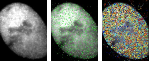
We are also interested in DNA repair and chromosome maintenance in eukaryotes, which we explore in collaborations with different labs. This image shows localisations of individual cohesin (Scc1) proteins that are bound to DNA (in green) and the tracks of molecules diffusing in the nucleoplasm (in rainbow colours).
Find out more about our study on the cohesin loader protein Scc2/Nipbl with the Nasmyth lab: Rhodes et al. eLife 2017.
With the Brockdorff lab, we investigated how the chromatin regulator SMCHD1 is recruited to specific regions of the genome, identifying a loading factor and ATPase activity that govern its selective targeting: Constantinescu et al. Nature Communications 2026.